MEDICAL
COSMETIC
Get In Touch
Phone (medical)
(403) 457-1900
Phone (cosmetic)
(403) 202-4038
Address
Business Hours
- Mon - Fri
- -
- Sat - Sun
- Closed
MEDICAL
COSMETIC
MOLE CHECK / SKIN CANCER & SURVEILLANCE
Dr. Alanen is an authentic expert in the diagnosis and treatment of skin disease. He is a recognized skin cancer specialist, having performed over 14,000 surgeries (including Mohs' surgeries).
What Is A Mole?
The term “mole” includes all major types of brown spots – nevus, keratosis, and lentigo. Even for a dermatologist, it can be difficult to distinguish nevus from keratosis and lentigo. This dilemma is confronted by the Dermatologist performing a biopsy. Theoretically, only nevus can turn into melanoma.
How Does Mole Mapping Work?
How Does Mole Mapping Work?
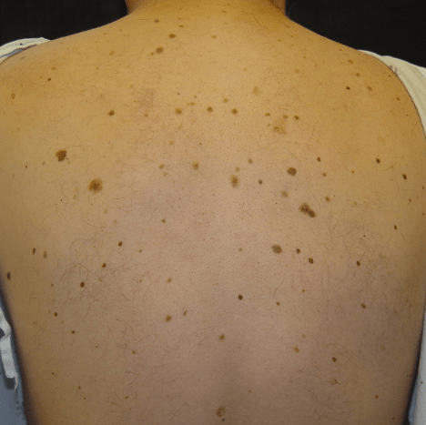
Dysplastic Nevi – Precursor To Melanoma
Most moles or nevi, are innocent biologically, however, some may transform into melanoma, the most serious type of skin cancer. Dysplastic Nevi, often referred to as atypical moles or atypical nevi, are regarded as potential precursors of malignant melanoma, the most aggressive form of skin cancer.
It is thought that only a small proportion of dysplastic nevi become melanoma. The greater the number of these lesions that a patient has, the greater should the melanoma risk; those who have ten or more have more than ten times the risk of developing malignant melanoma as compared with the general population.
Moles which show concerning features are often removed so as to prevent melanoma and also to have confirmation from the laboratory after tissue is examination by a pathologist.
Mole Mapping For Early Detection
Up to fifteen percent of the Canadians have these moles. At times it is difficult to distinguish dysplastic nevi from early melanoma. Often, a handheld magnification device called a dermatoscope is used; this device helps to visualize subsurface aspects of the mole.
When concerning features are noted, the mole is removed so that it can be examined under the microscope and the definitive diagnosis can be established. Patients with numerous dysplastic nevi are at high risk of developing malignant melanoma. Mole mapping technology allows for early melanoma detection; read more here about mole mapping.
ABCDE Criteria For Self Checking Moles
Our team understands that our patients are unique and have very different cosmetic goals. We strive to help you achieve the best results and will customize a 3D Personalized Laser Procedure plan in the initial consultation.
While many people have been able to notice an improvement in their skin quality after a single treatment, it may require a few sessions to see desired results. We generally recommend 2-3 follow up treatments spaced a month or two apart, however, results will vary from person to person.
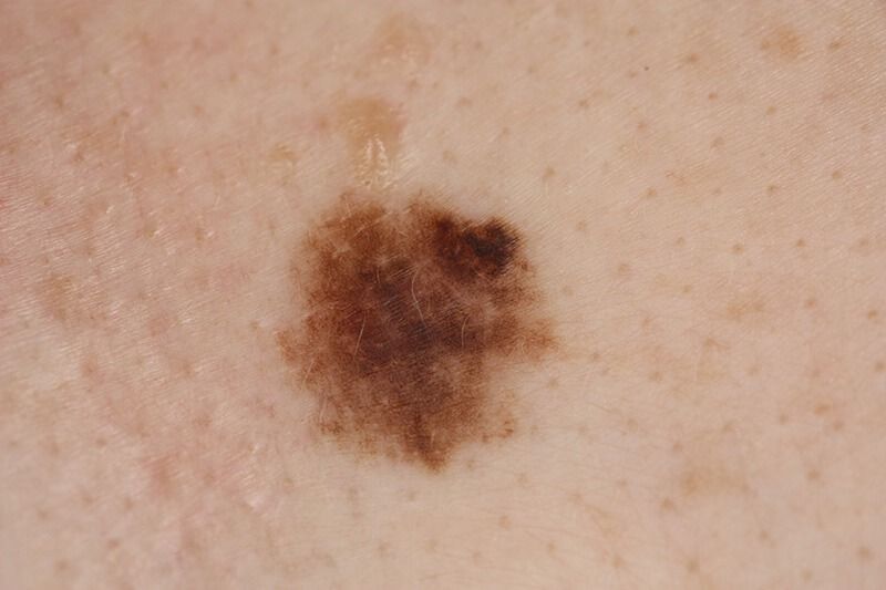
- Asymmetrical shape. Look for moles with irregular shapes
- Border. Look for moles with irregular, notched, “saw-toothed” borders.
- Colour irregularity. Look for moles that have multiple shades of brown or an uneven distribution of colour.
- Diameter. Most melanoma cancers are more than 6 mm in maximum diameter, although exceptions exist.
- Evolution or New Elevation. This is the MOST important criterion. Look for changes over time, such as a mole that grows in size or that changes colour or shape.
Mole Mapping is the best method to follow change or evolution of your moles.
How Does Mole Mapping Work?
When you arrive, you are greeted by our experienced and trained staff. We recognize that you are busy and every effort is made to be on time. High resolution digital images of your moles are taken in standardized poses.
This requires removal of clothing. We do not do genital photography, however. If a genital mole is of concern, this is examined clinically by the dermatologist. We pay particular attention to moles that you are worried about, however, it is noteworthy that many of the concerning lesions that we identify may not be the same moles that are of concern to the patient.
Moles of the scalp are difficult to photograph because of hair, of course, and a clinical examination is
required for assessment in this area of the body.
Do I Need A Referral?
Referrals from general practitioners are needed or you can
book for a private consultation.
How Long Does The Procedure Take?
This depends on the number of moles that you have but start to finish is classically less than an hour of your time, unless there are a very large number of moles.
Who Performs My Mole Mapping ?
Nurses or other trained health professionals with experience in dermatology do the image capture. The mole map images are subsequently assessed by dermatologist Dr. Kenneth Alanen who has subspecialty interest and expertise in moles and melanoma. Dr. Alanen’s assessment is completed within one week of your visit.
When Do I Get Seen In Follow-Up?
This depends on your individual risk factors and the number and type of moles that you have. For most patients, it is between six and twelve months.
Does It Hurt?
No, it is not painful. It is a completely non-invasive procedure.
What If A Concerning Lesion Is Found?
You will be contacted and it will be recommended that a biopsy or removal of a suspicious lesion should be performed. If feasible, a biopsy or removal may be done on the same day.
Please remember that not all suspicious lesions turn out to be melanoma. Concerning lesions identified during a mole mapping session are removed in Dr. Alanen’s dermatology clinic Derm.ca.
Will This Always Detect Melanoma?
Every effort is made for early detection of melanoma. No technology or amount of medical training is 100% effective for early detection, but this approach (authentic formal dermatology training, significant clinical experience, cutting edge technology), represents the highest level of diagnostic expertise.
Schedule A Consultation
Thank you for your interest in our clinic.
Medical Dermatology appointments require a referral from your Family Doctor or General Practitioner, while Cosmetic procedures do not.
A fee of $100 will be charged should you choose not to obtain a referral for any Medical appointments. Please contact us at 403-457-1900 or email us at info@derm.ca if you are unclear as to whether your issue is Medical or Cosmetic.
Also, you are welcome to use the acne.ca platform for secure electronic dermatology consultations.
HERE’S WHAT OUR PATIENTS ARE SAYING…
“I had a number of suspicious looking moles and other brown marks on my back, chest, and shoulders which were very concerning. I saw Dr. Alanen and cannot express how knowledgeable and thorough he was in going through the Mole Mapping process. He explained everything and answered all of my questions. Luckily there was nothing to be concerned about but I now have a baseline to work from. I highly recommend Derm.ca and Dr. Alanen”
-Gilbert L.
News
Mon: Cosmetic Appointments 8:00am – 4:45pm
Sat: Appointment Only (Cosmetic)
Sun: CLOSED
Holidays: CLOSED
Connect With Us
All Rights Reserved | Derm.ca & Derm.ca


