Calgary Skin Cancer Clinic
Comprehensive skin cancer care in one trusted Calgary dermatology clinic—from full-body skin examinations and advanced digital mole mapping to biopsies, diagnosis and skin cancer surgery.
Led by Dr. Ken Alanen, board-certified dermatologist and triple-certified surgical dermatologist, Derm.ca provides experienced, efficient and safe skin cancer care.
Early Skin Cancer and Melanoma Detection in Calgary
Early detection can save lives.
When melanoma is detected early, before it has spread, the five-year relative survival rate is approximately 97% or higher. Once melanoma spreads to distant areas of the body, survival decreases dramatically. This is why regular skin examinations, monitoring changing moles and promptly assessing suspicious lesions are so important.
At Derm.ca, patients can access comprehensive skin cancer screening, mole assessment and ongoing surveillance at our Calgary dermatology clinic. Dr Alanen is the most experienced skin cancer surgeon in Calgary.
The ABCDE Approach
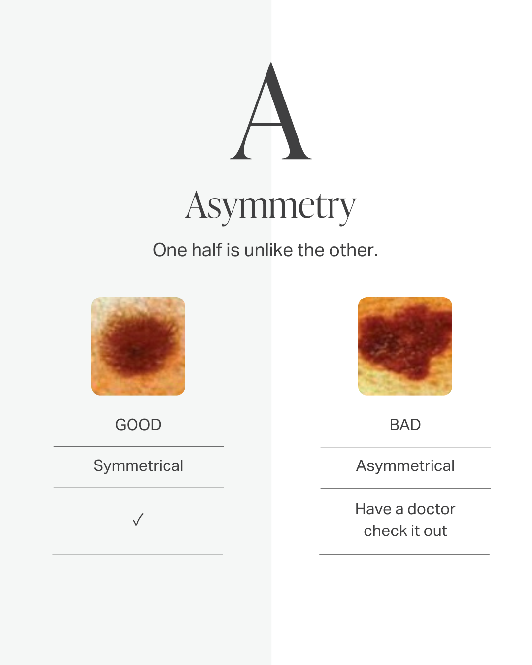
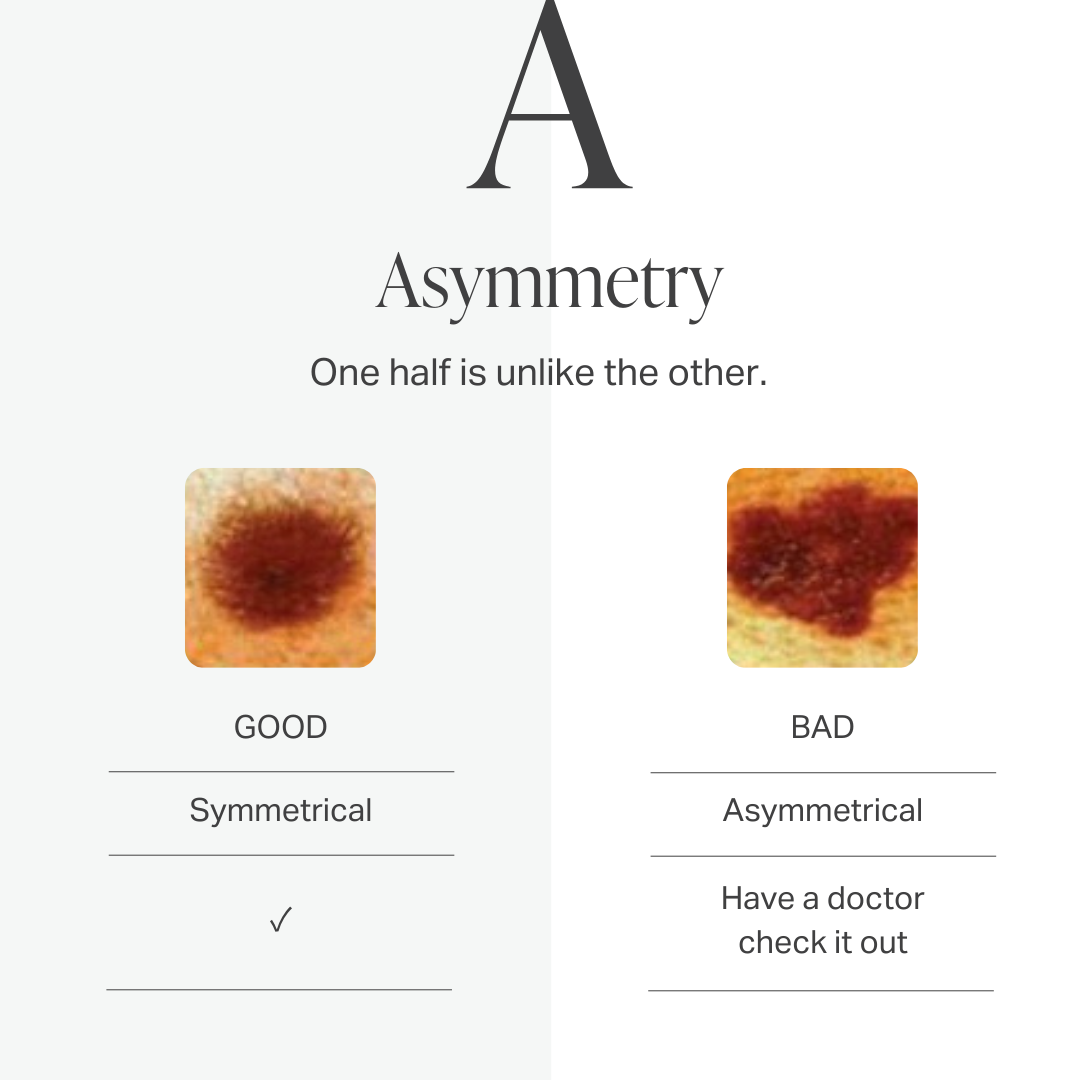
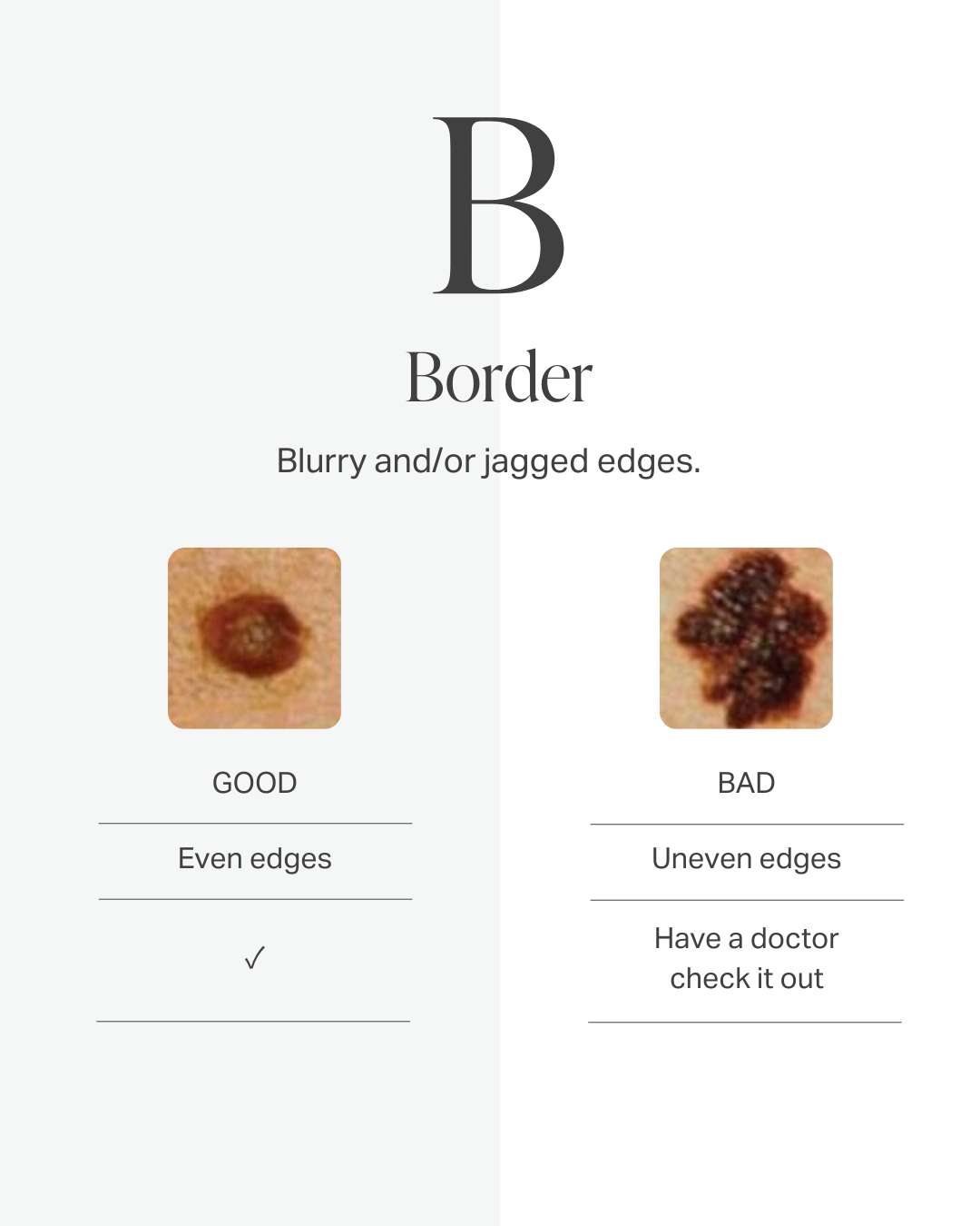
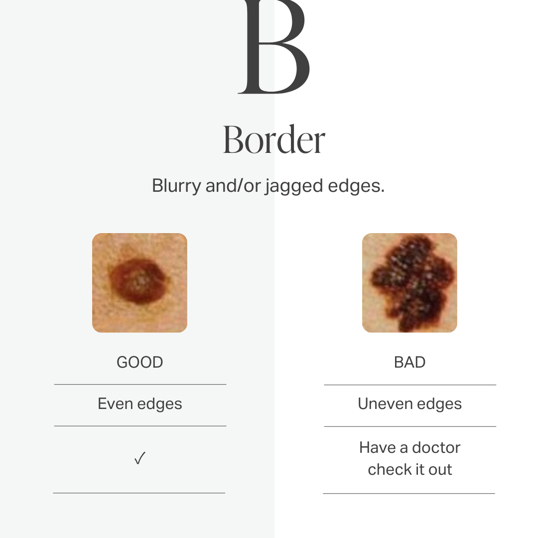
The "ABCDE" approach is a useful way to follow your moles. Be aware, however, that many serious moles may not violate these criteria. Also, many moles do indeed violate these rules but are biologically innocent. Have your skin checked by an authentic expert dermatologist.
Types of Cancers + Conditions
Actinic Keratoses
Actinic Keratoses (AK) are potential precancerous skin lesions. They occur as a consequence of cumulative low-grade sun exposure over many years.
Actinic Keratoses form not just from intentional suntanning but from every day casual sunshine such as walking to and from your car, a brief stroll with your dog, a walk with friends through the neighborhood, and even exposure to the sun through the windows in your car.
Actinic Keratoses can occur anywhere on the body, however, they are most commonly found on the face, neck, forearms, backs of the hand, any other sun-exposed parts of the body.
AKs look like a patch of dry skin, and treatment of AKs is necessary as they commonly transform into squamous cell carcinoma.
The treatment options are varied but include liquid nitrogen, surgical excision, topical medications (5-fluorouracil, imiquimod) as well as medium-deep skin peels using trichloroacetic acid, and photodynamic therapy.
If you think you may have Actinic Keratoses, we strongly recommend that you schedule an appointment with Dr. Alanen immediately.
Basal Cell Carcinoma
Basal Cell Carcinoma (BCC) is the most common type of cancer found in humans. It occurs most often on the face, neck, arms, and hands. As with Squamous Cell Carcinoma (SCC), the most important risk factor for Basal Cell Carcinoma is years of exposure to ultraviolet light.
Although not necessarily directly related to the use of tanning beds, tanning beds in conjunction with a history of phototherapy for psoriasis are considered significant risk factors for Basal Cell Carcinoma.
BCC most often looks like a “non-healing sore” or persistent pimple. However, in some cases, it may resemble a scar.
Treatment
Basal Cell Carcinoma is best treated by surgical removal.
Dr. Alanen’s Expertise
The microscopic examination and diagnosis part of the procedure is often regarded in the dermatology community as the most difficult part of the procedure. However, Dr. Alanen is a recognized subspecialist in microscopic diagnosis of skin cancer.
Squamous Cell Carcinoma
Squamous Cell Carcinoma (SCC) is the second most common type of skin cancer in humans. It typically develops on sun-exposed areas of the body, including the face, ears, lips, hands, arms, and legs. Like Basal Cell Carcinoma (BCC), the primary risk factor for Squamous Cell Carcinoma is cumulative ultraviolet light exposure over many years.
Additional risk factors include a history of severe sunburns, fair skin, immunosuppression, and exposure to certain chemicals or radiation. Individuals with a history of actinic keratoses (precancerous skin lesions) are also at increased risk for developing SCC.
SCC commonly appears as a firm, red nodule or a flat lesion with a scaly, crusted surface. It may also present as an open sore that doesn't heal, or a wart-like growth. Unlike BCC, SCC has the potential to spread to other parts of the body if left untreated, making early detection and treatment crucial.
Treatment
Squamous Cell Carcinoma is most effectively treated through surgical removal.
Dr. Alanen's Expertise
The microscopic interpretation component of Mohs' surgery requires extensive specialized training and is considered one of the most challenging aspects of the procedure. Dr. Alanen's recognized expertise as a subspecialist in the microscopic diagnosis of skin cancer ensures accurate identification and complete removal of SCC while minimizing the risk of recurrence.
Dysplastic Nevi
Dysplastic Nevi, also referred to as atypical moles or atypical nevi, are potential precursors of malignant melanoma, the most aggressive form of skin cancer.
Only a small proportion of dysplastic nevi transform into melanoma The higher the number of these moles someone has, the higher the risk; those who have 10 or more have 12 times the risk of developing melanoma as compared with the general population.
Medical reports indicate that about 2 to 8 percent of the Caucasian population have these moles. At times it is difficult to distinguish dysplastic nevi and early melanomas.
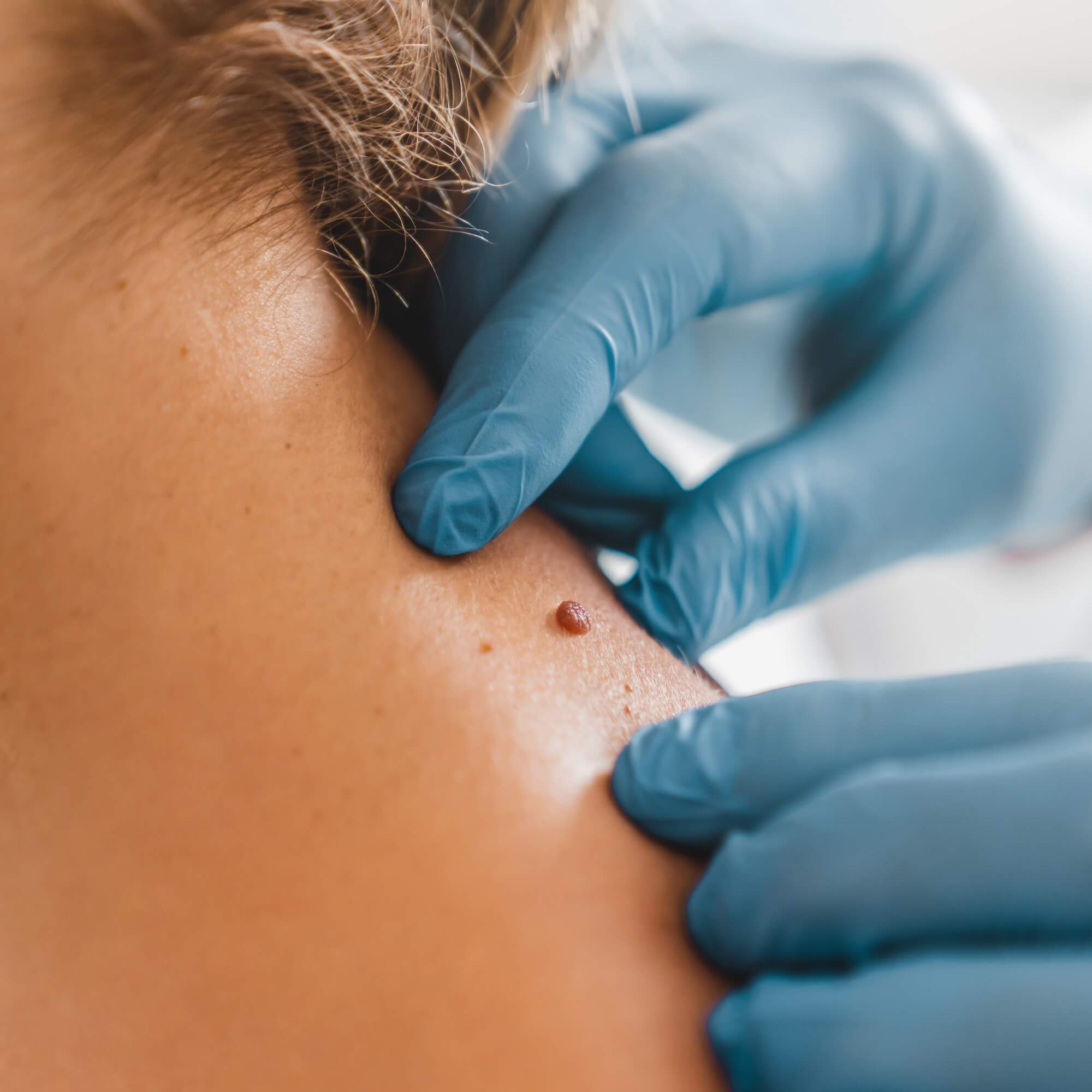
Often, a handheld magnification device called a dermatoscope is used. When concerning features are noted, the mole is removed so that it can be examined under the microscope and the definitive diagnosis can be established.
Patients with numerous dysplastic nevi are at high risk of developing malignant melanoma. Mole mapping technology allows for early melanoma detection; read more here.
If you have a concerning mole, please book an appointment.
Melanoma
Melanoma is much less common than some other types of skin cancers. But melanoma is more dangerous because it’s much more likely to spread to other parts of the body if not caught and treated early.
Malignant melanoma is the most serious type of skin cancer. It may start from a mole or arise on normal skin. If melanoma is detected early, it is classically curable. However, if melanoma spreads from the skin to lymph glands and other organs, it is often fatal.
The risk factors for melanoma include: fair skin, reddish hair, numerous moles, numerous freckles, a history of sunburns (particularly in youth), a personal or family history of numerous atypical (dysplastic) moles.
The A, B, C, D, E, Identification Approach
To help you identify characteristics of unusual moles that may indicate melanomas or other skin cancers, think of the letters A-B-C-D-E:
A. is for asymmetrical shape. Look for moles with irregular shapes, such as two very different-looking halves.
B. is for irregular border. Look for moles with irregular, notched or scalloped borders characteristics of melanomas.
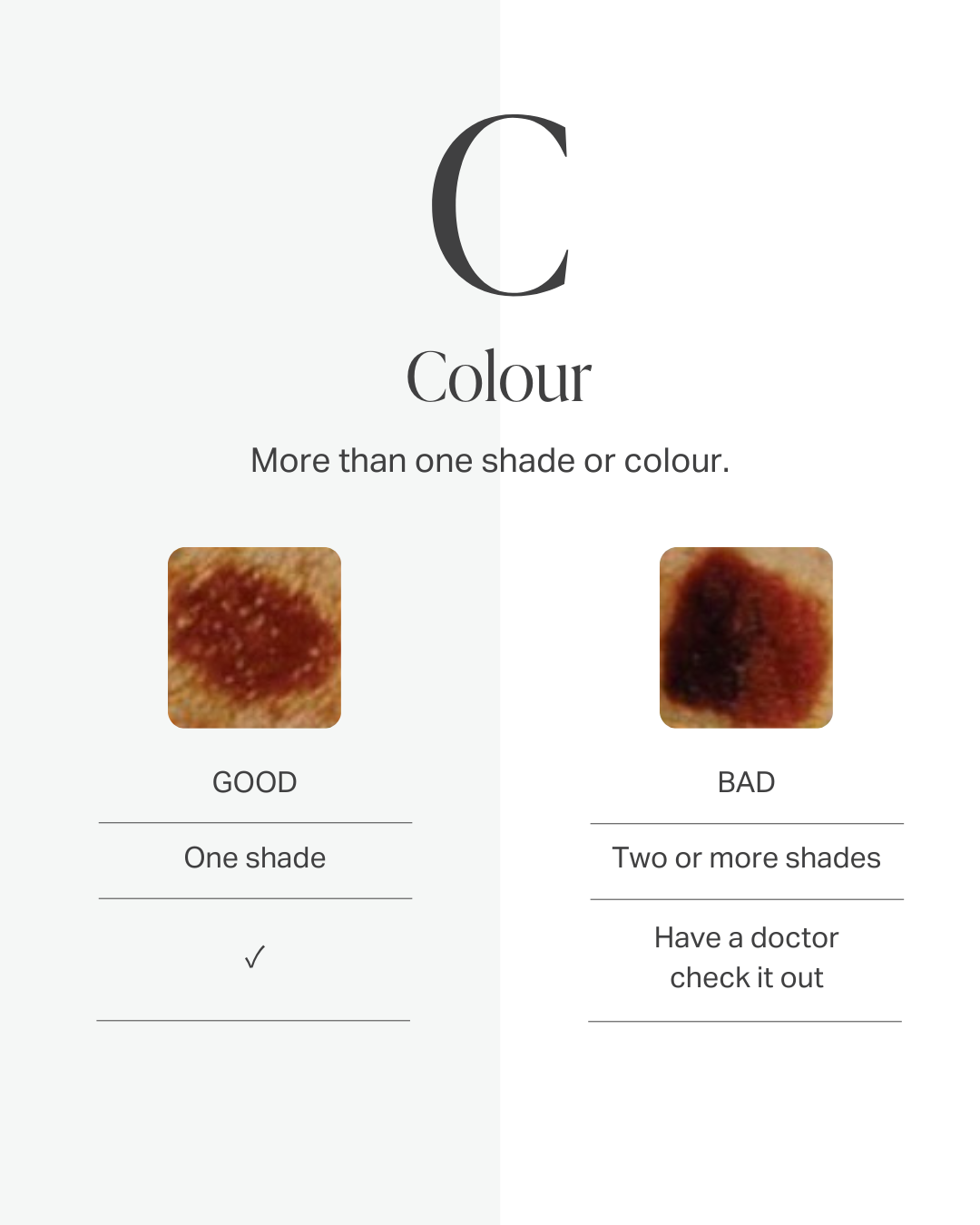
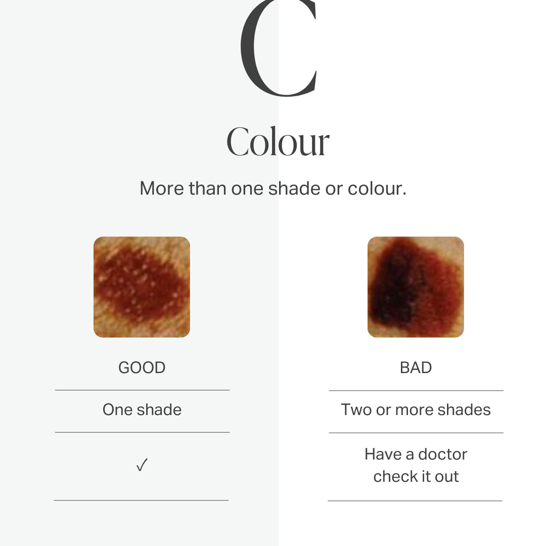
C. is for changes in color. Look for growths that have many colors or an uneven distribution of color.
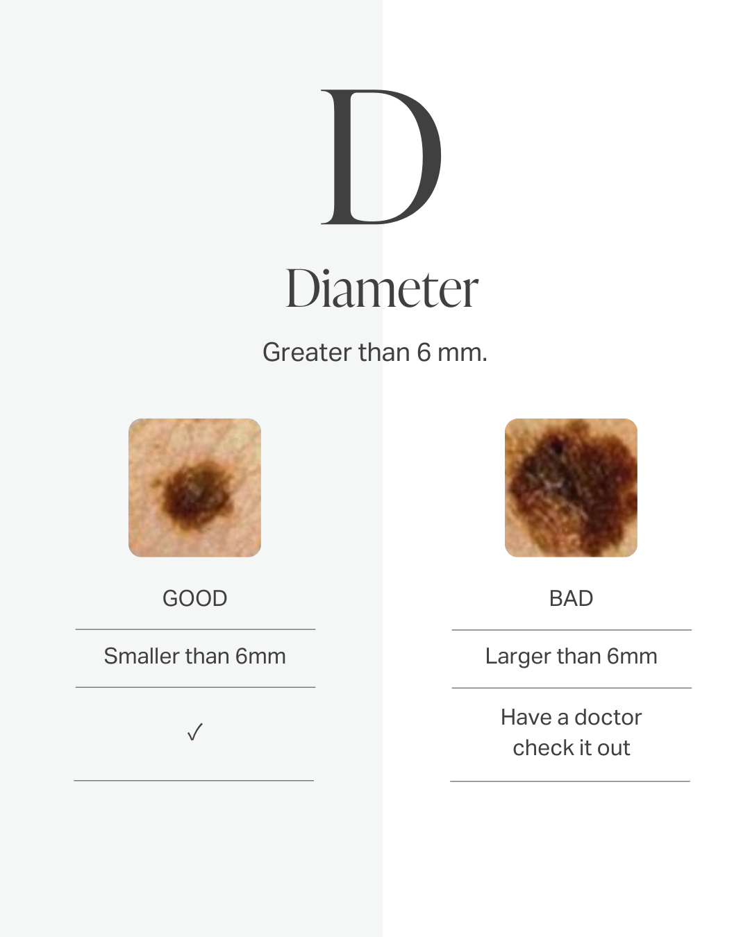
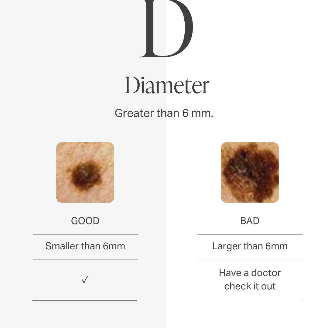
D. is for diameter. Look for new growth in a mole larger than 1/4 inch (about 6 millimeters).
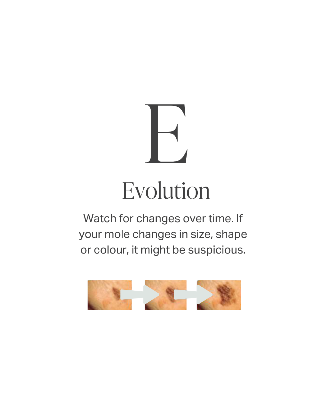
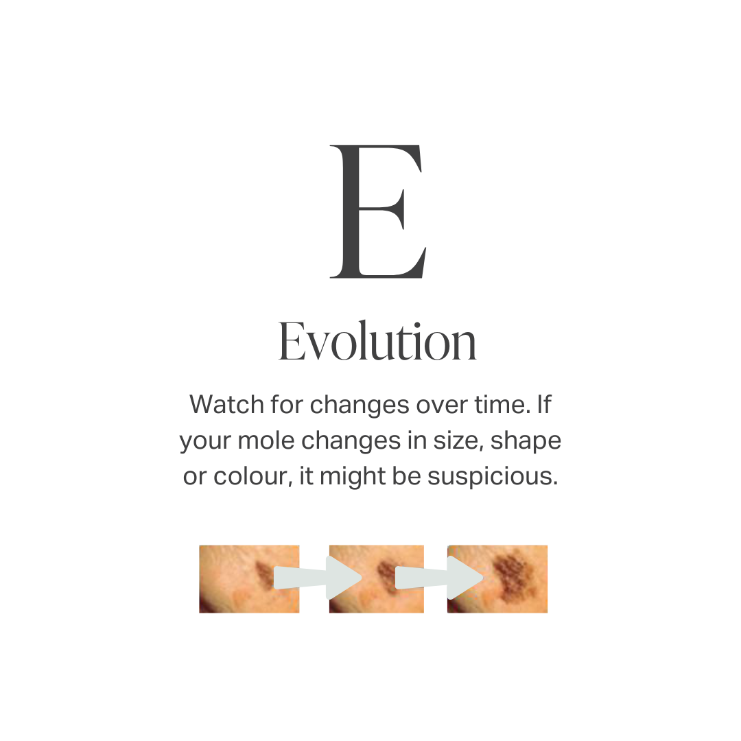
E. is for evolving. Look for changes over time, such as a mole that grows in size or that changes color or shape. Moles may also evolve to develop new signs and symptoms, such as new itchiness or bleeding.
Complete Skin Review: Board certified Dermatologist-Led Mole Checks and Skin Cancer Screening
Your skin cancer assessment may include a review of your medical and family history, a full-body skin examination, dermoscopic evaluation and personalized recommendations for monitoring suspicious or changing lesions.
If Dr. Alanen identifies an area of concern, a biopsy may be performed during the same appointment when medically appropriate. The tissue is then examined by a pathology laboratory to determine whether skin cancer is present.
This allows patients to move efficiently from screening to proper diagnoses to an appropriate treatment plan.
What happens during a Complete Skin Review?
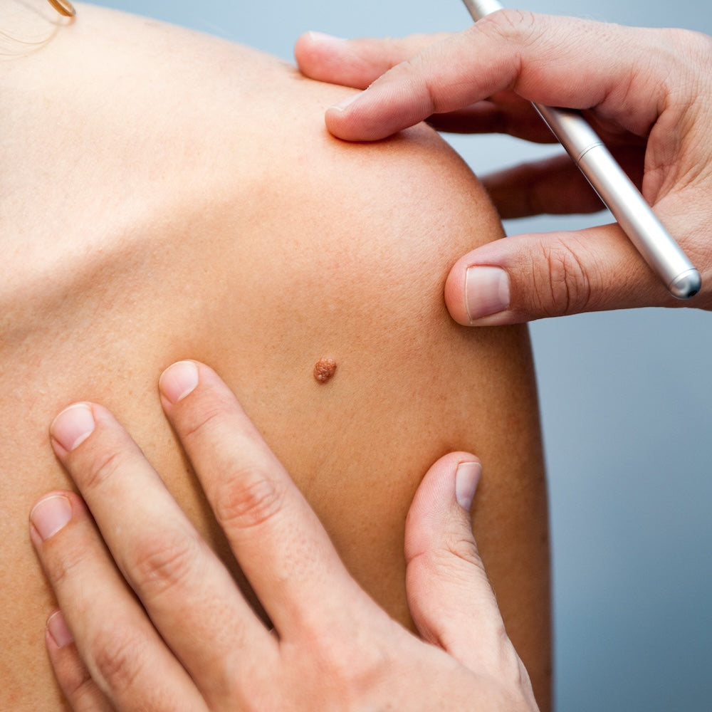
Dr. Alanen examines your skin paying particular attention for unusual moles, sun damage and precancerous sunspots. In some cases he may use a dermatoscope – a special dermatology developed device to magnify the skin and underlying structures to assist with the diagnosis.
If it is found that you have many moles, mole mapping may be recommended. If significant sun damage is noted on the face, Dr. Alanen may recommend subsurface high resolution computer analysis of your skin so as to quantify sun damage, blood vessel overgrowth, and pigmentation irregularities.
How long does a Complete Skin Review take?
Typically, Complete Skin Review takes only ten minutes or so.
Do I need a Complete Skin Review?
If you have a personal or family history of skin cancer, a history of sunburns or many suntans, fair skin, multiple moles, or fair hair, the answer is yes.
How reliable is a Complete Skin Review?
Very reliable. This relates to the intrinsic nature of dermatology being a visual pattern recognition – based specialty and sheer number of examinations that Dr. Alanen has performed over the years.
Will my concerns be addressed?
Yes.
What if a concerning spot is found?
Dr. Alanen may treat the lesion with cryotherapy (liquid nitrogen; “dry ice”) or remove (i.e. biopsy) the lesion.
Removal/biopsy can almost always be done on the same day as the Complete Skin Review — a very efficient system has been developed over the years that allows for this service.
Do you send tissue for pathology confirmation?
Yes. Every single time. The tissue is processed at the laboratory and mounted onto glass slides so that it can be examined and diagnosed under the microscope by the pathologist. Of note, Dr. Alanen is also a board-certified pathologist as well as subspecialty certified dermatopathologist. If your report has an equivocal "confusing" diagnosis, Dr. Alanen can review the specimen as part of his expert second opinion service.
Do you have an expert second opinion service?
No referral is needed.
Do I need a referral for a Complete Skin Review?
No referral is needed.
Is there a cost for Complete Skin Review?
A non-referred visit with dermoscopy examination is not covered by provincial insurance plans. A Complete Skin Review costs $300.
Advanced Digital Mole Mapping and Full-Body Skin Screening
Digital mole mapping is an advanced skin-imaging service that creates detailed photographs of the skin and a baseline record of your moles. These images can be compared over time to help identify new or changing lesions that may require closer examination. Usually Dr Alanen will determinate based on the high risk patients howe often they need to have the Mole Mapping done.
One Calgary Clinic for Skin Cancer Detection and Treatment
For cases that can be treated within our clinic, patients can receive their skin examination, mole mapping, biopsy, diagnosis and skin cancer surgery through one experienced dermatology team.
There is no need to search for separate private providers for each stage of care. Your assessment, results and treatment plan are coordinated by the Derm.ca team, helping make the process more efficient, reassuring and easier to navigate.
If a condition requires specialized hospital-based or multidisciplinary care, we will explain the next steps and coordinate the appropriate referral.

Skin Biopsies and Skin Cancer Diagnosis
A biopsy is the medical procedure used to collect a small tissue sample from a suspicious skin lesion. The sample is assessed by a pathology laboratory to confirm whether the lesion is benign, precancerous or cancerous.
Depending on the clinical findings and pathology results, our team will explain the diagnosis, recommended treatment and next steps clearly and compassionately with each patient.
Skin Cancer Surgery in Our Calgary Clinic
When skin cancer is confirmed and can be appropriately treated at Derm.ca, surgery can often be arranged promptly within our Calgary dermatology clinic at Derm.ca
Dr. Alanen daily performs skin cancer surgery and has completed more than 40,000 skin cancer procedures. His extensive experience includes assessing and treating basal cell carcinoma, squamous cell carcinoma, melanoma and other suspicious or cancerous skin lesions.
Our goal is to provide safe, efficient, and carefully coordinated care at every stage—from early detection and diagnosis to surgical treatment, suture removal, and follow-up care to ensure each patient feels supported and satisfied with their outcome.
Although scarring is a potential risk following any surgical procedure, patients also have access to the expertise of Ildiko Juhasz, our experienced medical aesthetic laser clinician. Ildiko has treated surgical and other scars for decades and has performed scar-revision treatments for all Dr. Alanen’s patients for the past 18 years. She provides personalized care designed to improve the appearance, texture, and healing of post-surgical scars and help each patient achieve the best possible result.